In The Figure Which Diagram Of A Cell Wall Is A Gram Negative Cell Wall
B it is sensitive to lysozyme. D it contains teichoic acids.

In figure 43 which diagram of a cell wall is a toxic cell wall smaller gram negative in figure 43 which diagram of a cell wall has a wall that protects against osmotic lysis.

In the figure which diagram of a cell wall is a gram negative cell wall. 27 in figure 43 which diagram of a cell wall is a gram negative cell wall. Need an extra hand. C both a and b in figure 43 which diagram of a cell wall is decolorized by acetone alcohol.
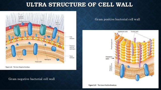
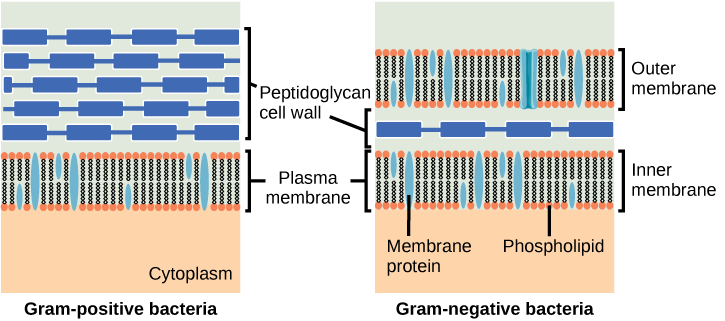
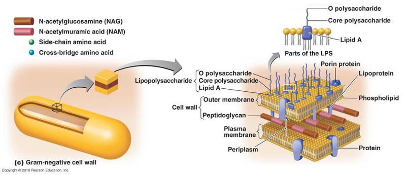
The bacterial cell wall consists of a polysaccharide complex called peptidoglycan. The cell wall is many times thicker in gram positive bacteria 3 100 nm thickness than in gram negative bacteria 3 8 nm fig. D neither a nor b.
The correct option is option b b the gram negative cell wall is figure b. Our entire perception of gram positive and gram negative walls ultimately relies on the response of bacteria to gram staining. Is part of a multicellular animal.
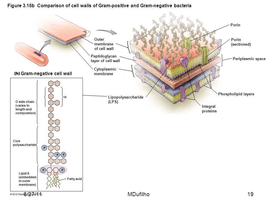
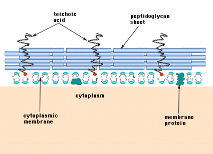
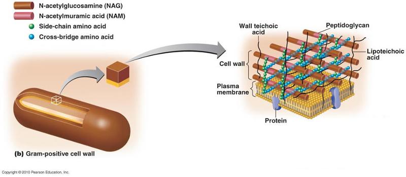
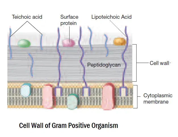
2 each of the following statements concerning the gram positive cell wall is true except a it maintains the shape of the cell. In gram positive bacteria peptidoglycan makes up as much as 90 of the thick cell wall enclosing the plasma membrane. The gram negative bacteria contain an outer membrane surrounding the cell wall.
See page 2 for a diagram of the gram negative cell wall and a video on gram staining. Has a peptidoglycan cell wall d. Together the plasma membrane and the cell wall outer membrane peptidoglycan layer and periplasm constitute the gram negative envelope 5 9.
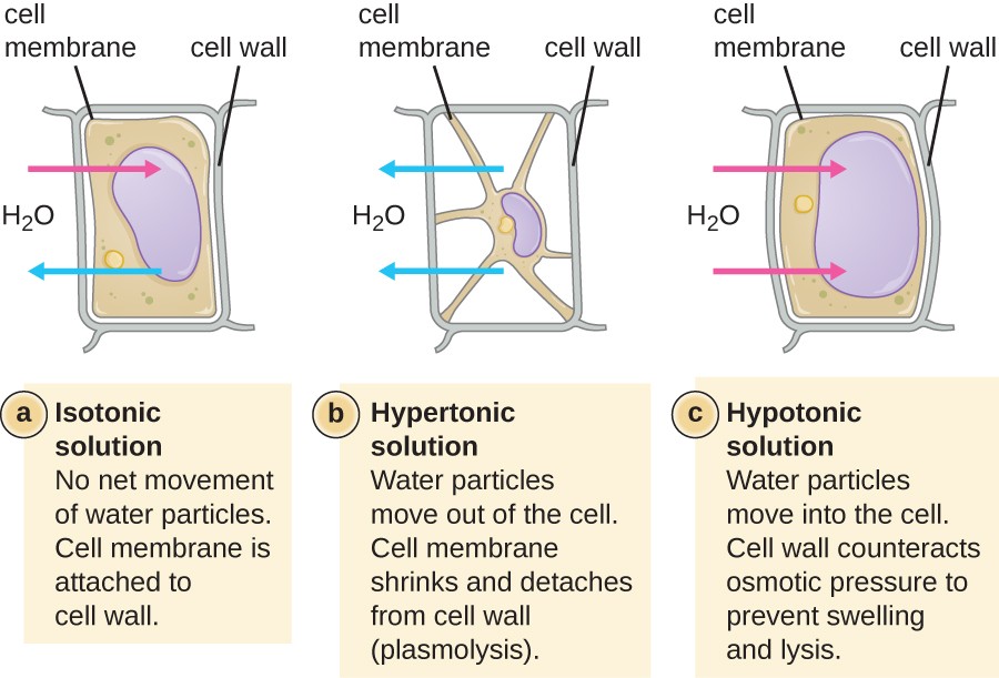
C it protects the cell in a hypertonic environment. The use of living organisms to make desired products. Is a plant cell.
Has a cellulose cell wall. Browse hundreds of biology tutors. C both a and b.
In figure 43 which diagram of a cell wall is a gram negative cell wall. In figure 43 which diagram of a cell wall has a wall that protects against osmotic lysis. You are observing a cell through a microscope note that it has no apparent nucleus you conclude that it most likely a.
E the answer cannot be determined based on the information provided. The main difference between gram positive and gram negative bacteria is that gram positive bacteria contain a thick peptidoglycan cell wall along with teichoic acid allowing the bacteria to stain in purple during gram staining whereas gram negative bacteria contain a thin peptidoglycan cell wall with no teichoic acid allowing the cell wall to. E it is sensitive to penicillin.
:max_bytes(150000):strip_icc()/plant_cell_wall_chloroplasts-5b64a485c9e77c0025a39acf.jpg) Cell Wall Structure And Function
Cell Wall Structure And Function
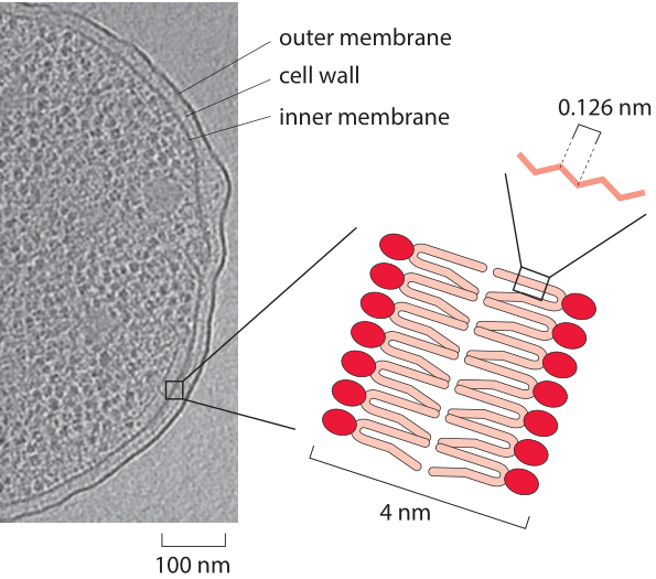
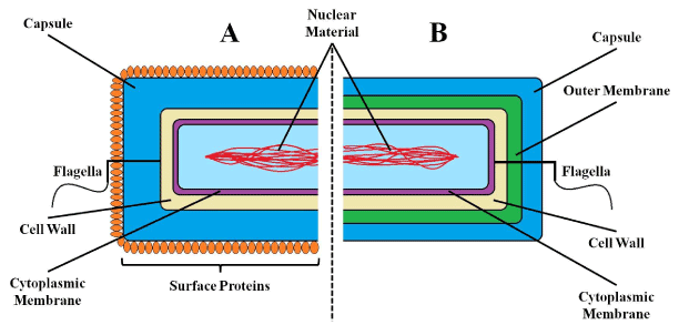 Figure 1

 Bacterial Cell Wall Synthesis
 Cell Structure And Function Ppt Video Online Download
 Schematic Views Of Gram Negative Top And Gram Positive Bottom
 Unique Characteristics Of Prokaryotic Cells Microbiology
 2
2
 Gram Negative Bacteria Wikipedia

 Peptidoglycans Sigma Aldrich
 13 1 Prokaryotic Diversity Diversity Of Microbes Fungi And By
 Diagram Of The Bacterial Cell Wall And The Respiratory Chain At The
 Structural Difference Between Gram Positive Bacteria And Gram
 Functional Anatomy Of Prokaryotic And Eukaryotic Cells Flashcards
 Unique Characteristics Of Prokaryotic Cells Microbiology
 Functional Anatomy Of Prokaryotic And Eukaryotic Cells Flashcards
Unique Characteristics Of Prokaryotic Cells Microbiology
 Differences Between Gram Positive And Gram Negative Bacteria
0 Response to "In The Figure Which Diagram Of A Cell Wall Is A Gram Negative Cell Wall"
Post a Comment