Identify Which Diagram Represents Cells That Produce And Circulate Cerebrospinal Fluid
The of a presynaptic neuron associates with the dendrite of a postsynaptic neuron. True or false a neuron is a collection of nerve fibers in the pns.
 Anatomy Lecture Exam 3 Set Flashcards Quizlet
Between 50 to 70 of csf is produced in the brain by modified ependymal cells in the choroid plexus and the remainder is formed around blood vessels and along ventricular walls.
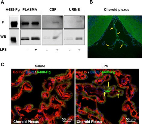
Identify which diagram represents cells that produce and circulate cerebrospinal fluid. Cerebrospinal fluid circulation begins with the pulsing of the choroid plexus. This is the site of communication between neurons. The structure located in the ventricles that produces cerebrospinal fluid is called the choroid plexus.
The choroid plexus consists of many capillaries separated from the ventricles by choroid epithelial cells. Identify which letter represents an oligodendrocyte. This protective device has many elements ranging from junctions between endothelial cells in the capillaries of.
This is clinically important because some drugs cannot penetrate the barrier. The traditional understanding of csf physiology assumes that 80 of csf is secreted by the choroid plexus into the ventricular cavities. Identify which diagram represents cells that produce and circulate cerebrospinal fluid.
It circulates from the lateral ventricles to the foramen of monro interventricular foramen third ventricle aqueduct of sylvius cerebral aqueduct. The cerebrospinal fluid is a clear protective fluid made by the cells of the choroid plexus and it is commonly abbreviated csf. Identify which diagram represents cells that produce and circulate cerebrospinal fluid.
Identify which letter represents an oligodendrocyte. Fluid filters through these cells from blood to become cerebrospinal fluid. Circulation of the cerebrospinal fluid.
It will eventually circulate throughout the subarachnoid spaces in the brain and spinal cord and then be absorbed into the bloodstream. A network of capillaries called the choroid plexus projects into each ventricle. Tiny cilia located on ependymal cells that also produce small amounts of csf help propel the fluid along.
Interposed between the blood and the csf by the endothelial cells of the capillaries and the choroid plexus. The ependymal cells maintain a bloodcsf barrier controlling the composition of the csf. The anatomy of the cerebrospinal fluid csf system includes the cerebral ventricles as well as the spinal and brain subarachnoid spaces cisterns and sulci.
The csf circulates from the. Identify which diagram represents a cell that produces a myelin sheath in the central nervous system. Additional openings in the fourth ventricle allow csf to flow into the subarachnoid space.
There is also much active transport of substances into and out of the csf as it is made. The ventricles and cerebrospinal fluid.
 Chiari I Malformation Syringomyelia Mayfield Brain Spine
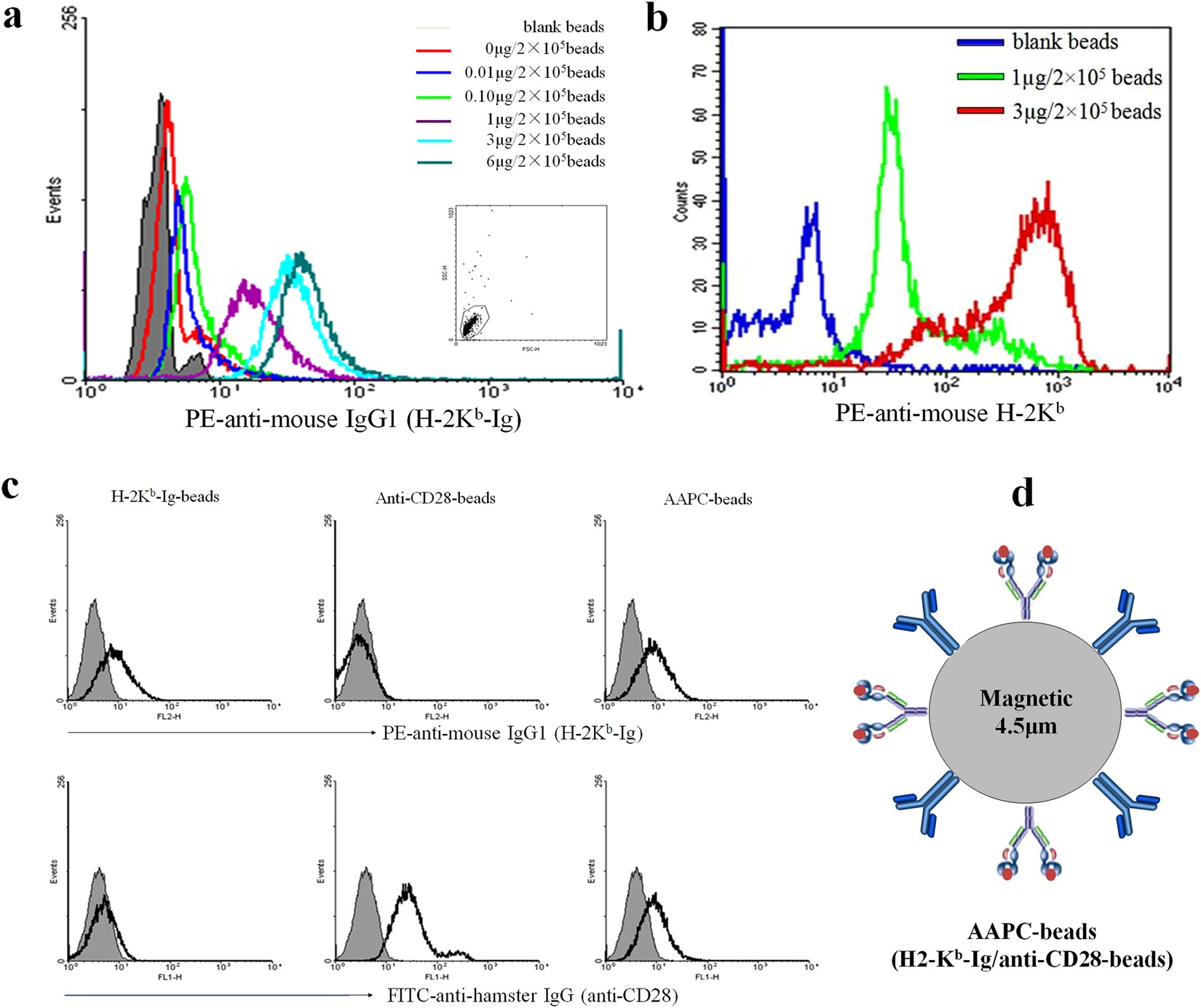 Frequency And Reactivity Of Antigen Specific T Cells Were
12 2 Nervous Tissue Anatomy Physiology
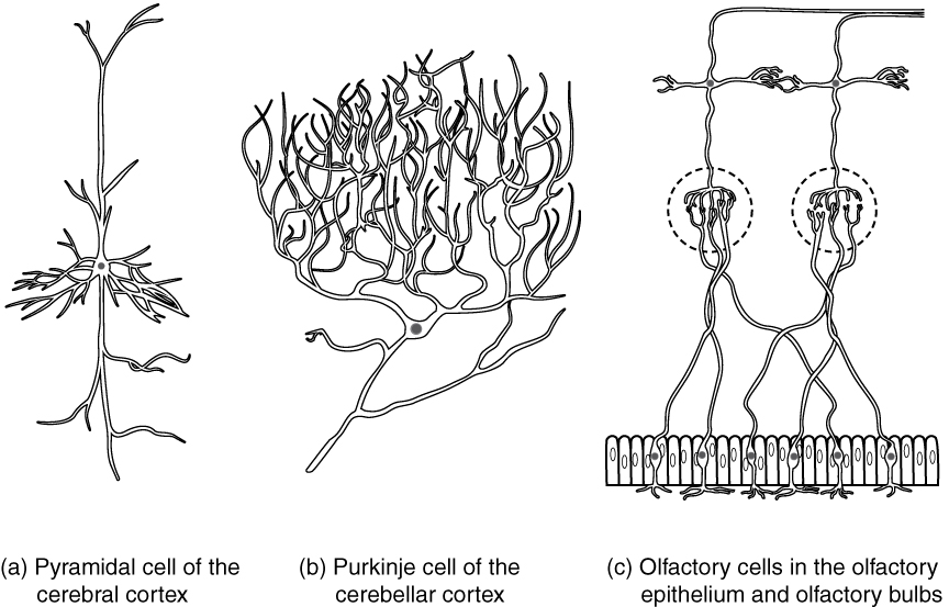 12 2 Nervous Tissue Anatomy And Physiology
 Plasminogen In Cerebrospinal Fluid Originates From Circulating Blood
14 2 Blood Flow The Meninges And Cerebrospinal Fluid Production And
 Development And Functions Of The Choroid Plexus Cerebrospinal Fluid
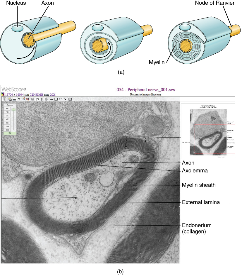 12 2 Nervous Tissue Anatomy And Physiology
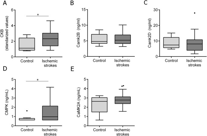 Characterization Of The Rat Cerebrospinal Fluid Proteome Following
 Cerebrospinal Fluid In The Brain Functions Production Video
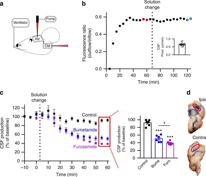 Cotransporter Mediated Water Transport Underlying Cerebrospinal
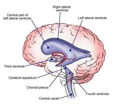 Ventricles Of The Brain Overview Gross Anatomy Microscopic Anatomy
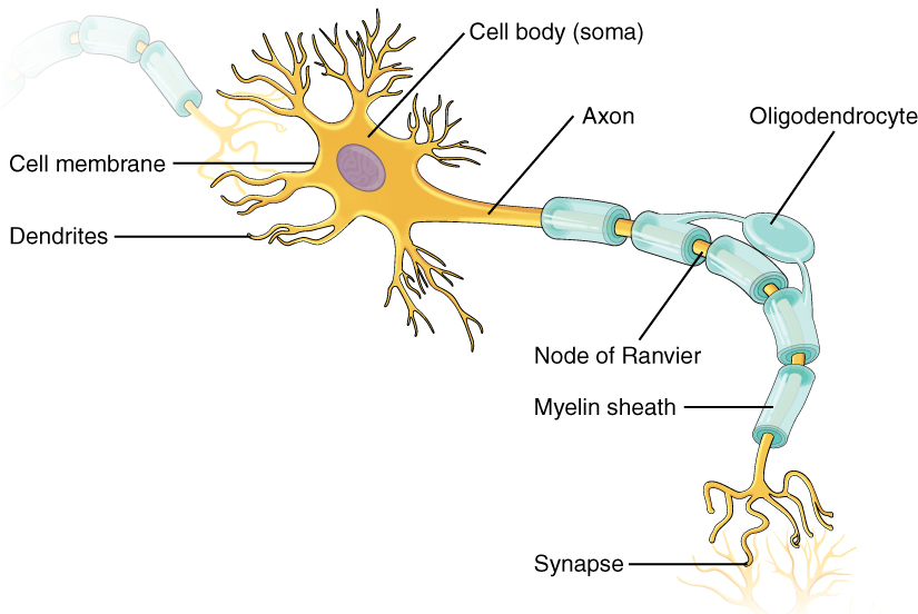 12 2 Nervous Tissue Anatomy And Physiology
 Intrathecal Dosing Leads To Measurable Cerebrospinal Fluid Csf
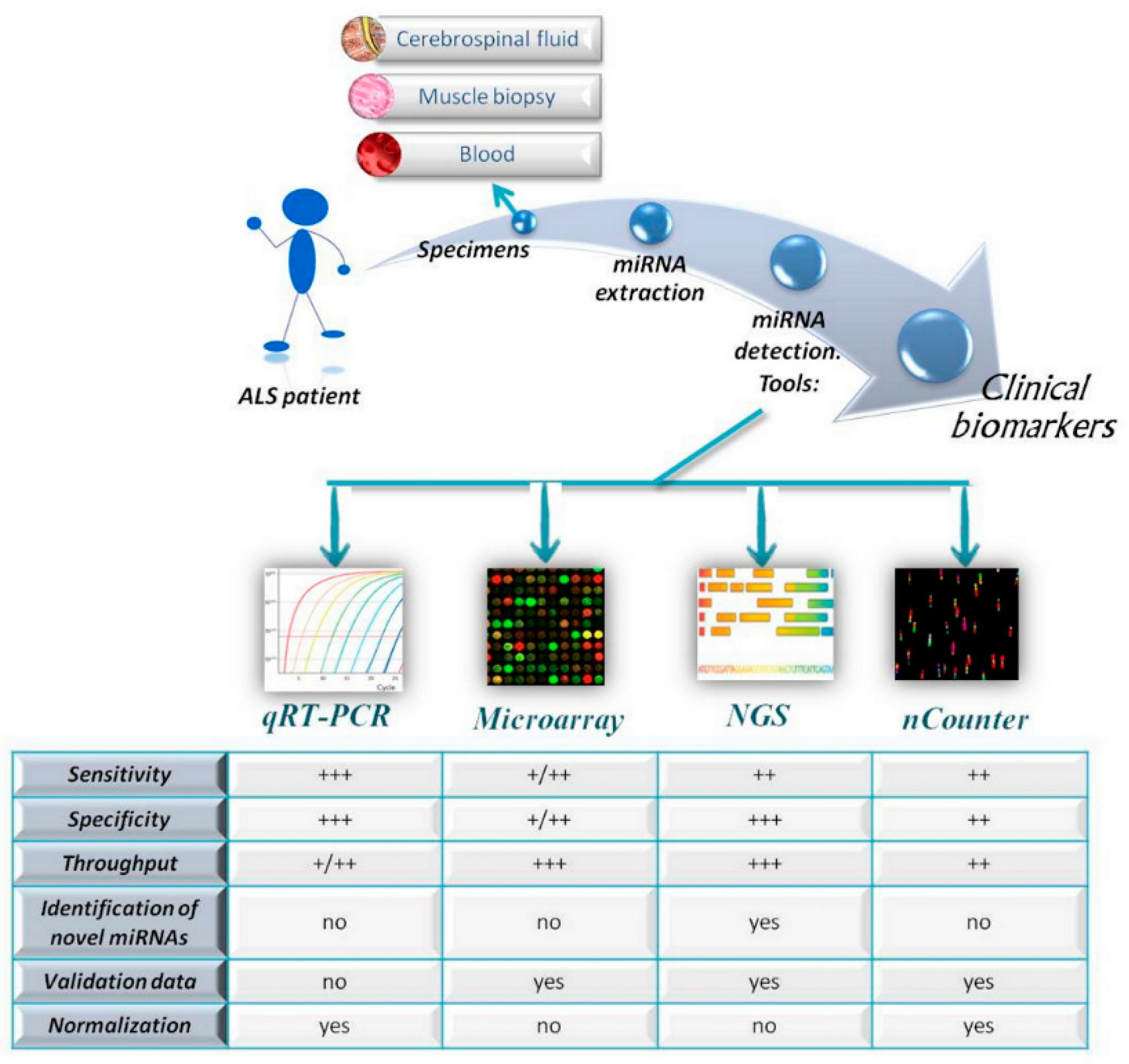 Cells Free Full Text Micrornas As Biomarkers In Amyotrophic
 Blood Cerebrospinal Fluid Barrier An Overview Sciencedirect Topics
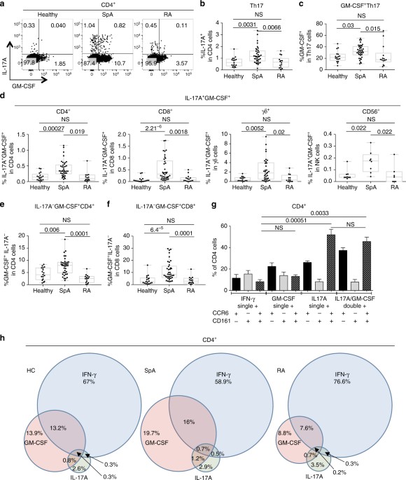 Unique Transcriptome Signatures And Gm Csf Expression In Lymphocytes

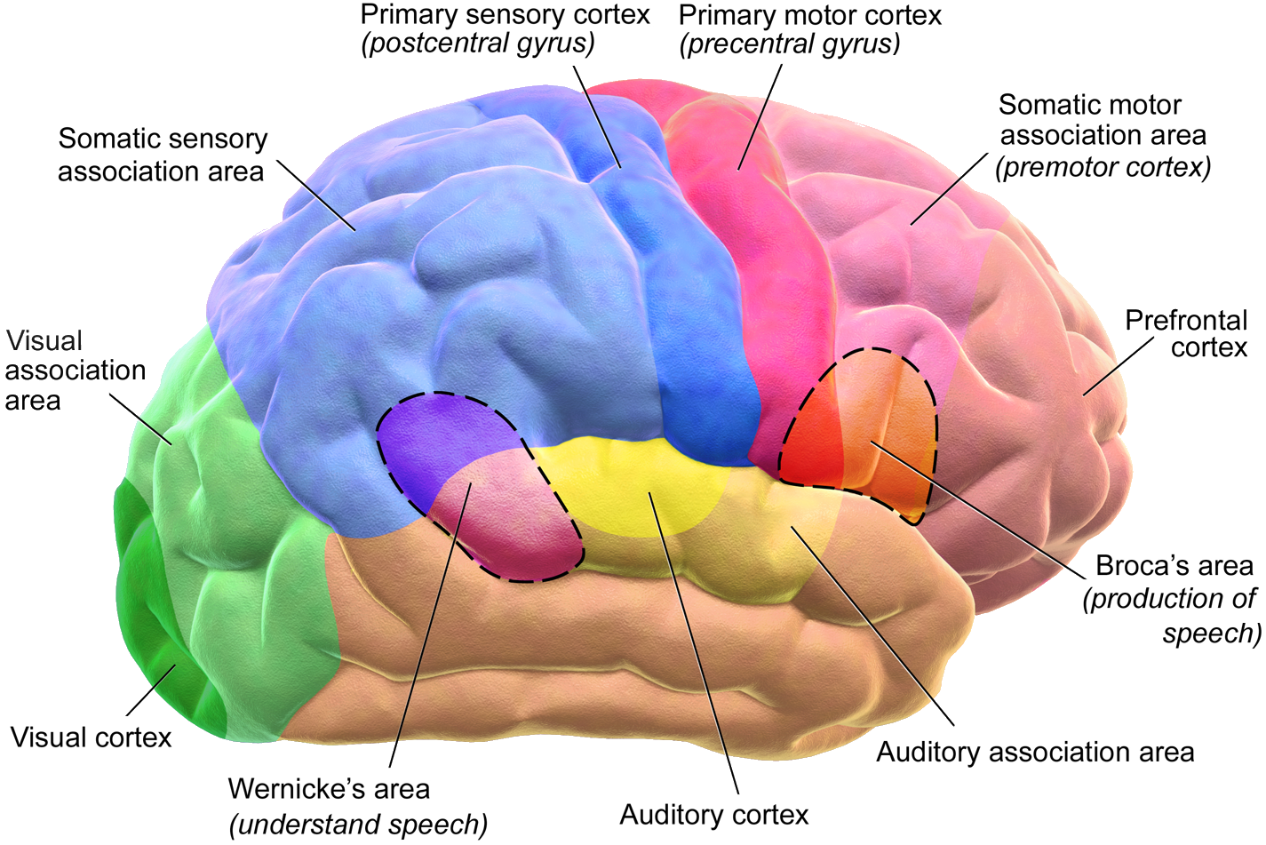
0 Response to "Identify Which Diagram Represents Cells That Produce And Circulate Cerebrospinal Fluid"
Post a Comment