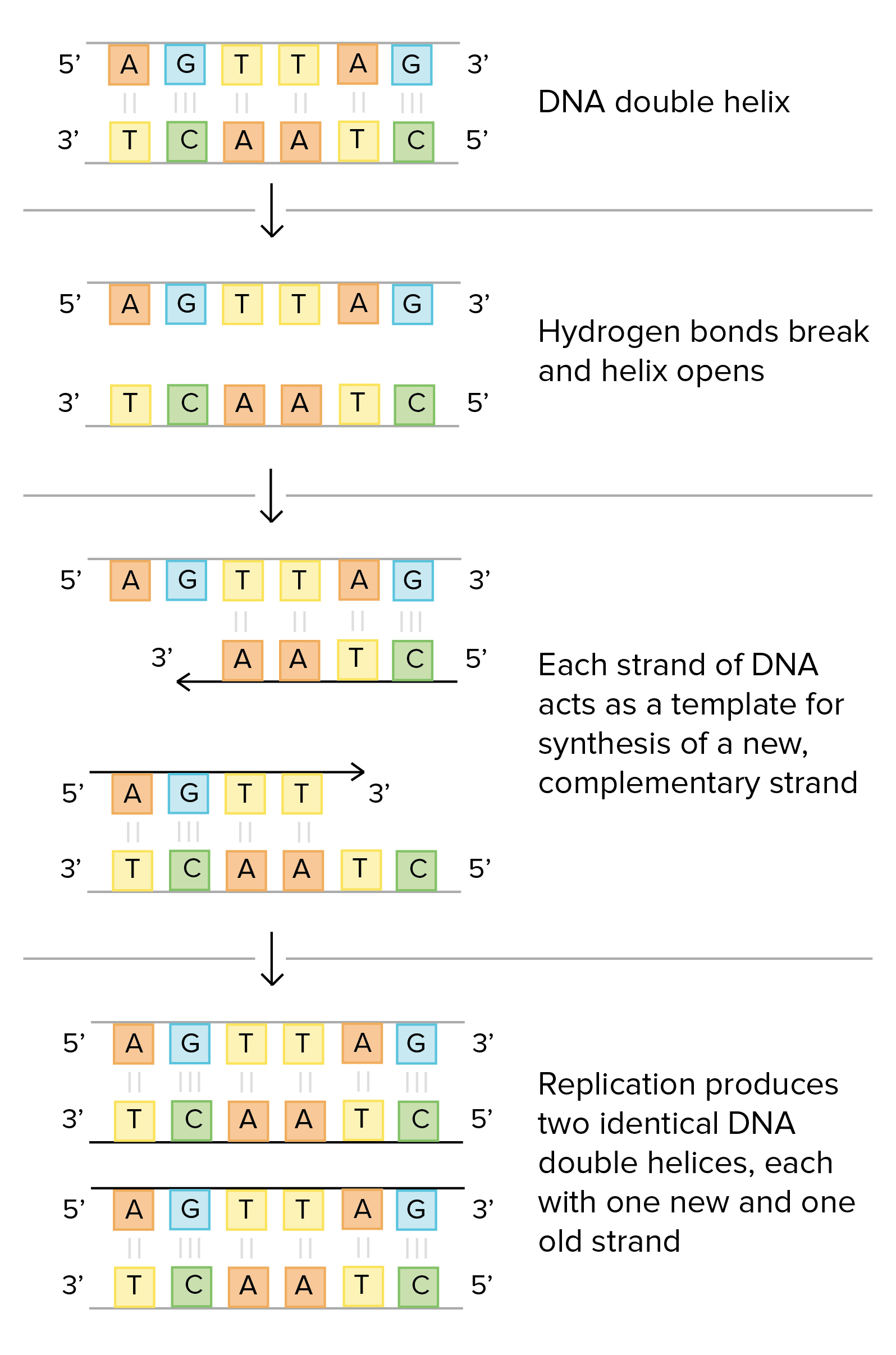
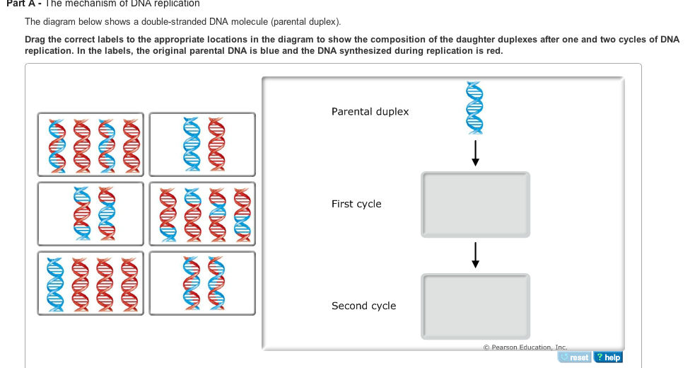
The Diagram Below Shows A Double Stranded Dna Molecule Parental Duplex
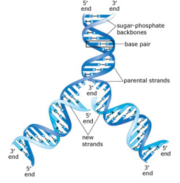
The structure of dna. In the labels the original parental dna is blue and the dna synthesized during replication is red.
 The Bacterial Dnaa Trio Replication Origin Element Specifies Single
In the labels the original parental dna is blue and the dna synthesized during replication is red.

The diagram below shows a double stranded dna molecule parental duplex. The dna molecule actually consists of two such chains that spiral around an imaginary axis to form a double helix spiral. In the labels the original parental dna is blue and the dna synthesized during replication is red. In the labels the original parental dna is blue and the dna synthesized during replication is red.
Drag the correct labels to the appropriate locations in the diagram to show the composition of the daughter duplexes after one and two cycles of dna replication. The parental dna is shown in dark blue the newly synthesized dna is light blue and the rna primers associated with each strand are red. The parental dna is shown in dark blue the newly synthesized dna is light blue and the rna primers associated with each strand are red.
The origin of replication is indicated by the black dots on the parental strands. Show transcribed image text the diagram below shows a bacterial replication fork and its principal proteins. Drag the correct labels to the appropriate locations in the diagram to show the composition of the daughter duplexes after one and two cycles of dna replication.
The diagram below shows a replication bubble with synthesis of the leading and lagging strands on both sides of the bubble. Drag the correct labels to the appropriate locations in the diagram to show the composition of the daughter dna molecules after one and two cycles of dna replication. Drag the correct labels to the appropriate locations in the diagram to show the composition of the daughter duplexes after one and two cycles of dna replication.
The diagram below shows a replication bubble with synthesis of the leading and lagging strands on both sides of the bubble. The diagram below shows a double stranded dna molecule parental duplex. Part a the mechanism of dna replication the diagram below shows a double stranded dna molecule parental dna.
The origin of replication is indicated by the black dots on the parental strands. The diagram below shows a double stranded dna molecule parental duplex. The diagram below shows a double stranded dna molecule parental duplex.
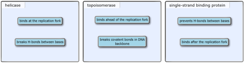
Drag the labels to their appropriate locations in the diagram to describe the name or function of each structure. Use pink labels for the pink targets and blue labels for the blue targets. Nucleic acids are made up of chains of many repeating units called nucleotides see bottom left of figure 1 below.
 Exam 3 Chs 5 Dna Structure And Replication Machinery 16 The

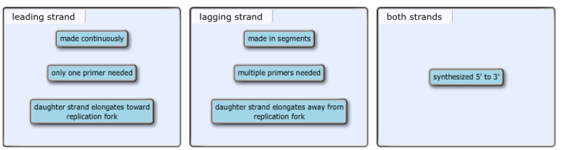
 Part A Comparing The Leading And Lagging Strands As The Two Parental
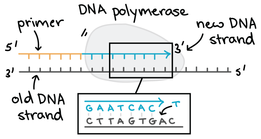 Molecular Mechanism Of Dna Replication Article Khan Academy
 Molecular Mechanism Of Dna Replication Article Khan Academy

 Exam 3 Chs 5 Dna Structure And Replication Machinery 16 The
 Mastering Biology Chapter 16 Rhs Homework
Working With Molecular Genetics Chapter 5 Dna Replication I V2 1
 Dna Replication Microbiology
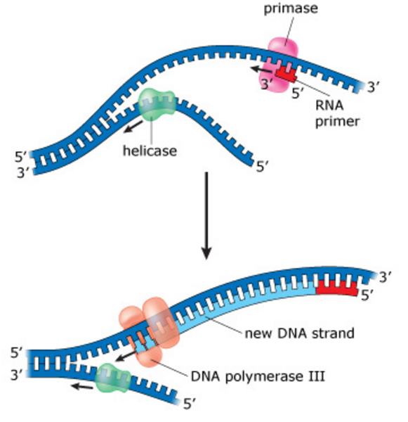
 The Diagram Below Shows A Bacterial Replication Fork And Its Principal Proteins Drag The Labels To Their Appropriate Locations In The Diagram To Describe The Name Or Function Of Each Structure Use

 An Introduction To Molecular Biology Replication Of Dna And Its
 Exam 3 Chs 5 Dna Structure And Replication Machinery 16 The
 Allolactose 27 Predict The Level Of Lac Operon Expression High
 Solved Dna Replication Is The Mechanism By Which Dna Is C
Dna Biology 1511 Biological Principles
Mastering Biology Chapter 16 Rhs Homework
 Exam 3 Chs 5 Dna Structure And Replication Machinery 16 The
0 Response to "The Diagram Below Shows A Double Stranded Dna Molecule Parental Duplex"
Post a Comment