In The Diagram Which Panel Shows Cells That Are In Interphase
As plant cells need to synthesis the cell wall during cytokinesis they do not create a cleavage furrow from the outside in cytokinesis starts where the spindle equator was and continues with new cell membrane and new cell wall material being laid down along this plate. Cholesterol 46 what structural components of the.
 In The Diagram Which Panel Shows Events Occurring During Anaphase
A a b b c c d d e e.
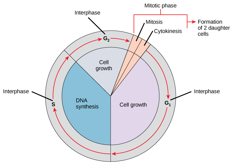
In the diagram which panel shows cells that are in interphase. Interphase is the stage of the cell cycle in which cells spend most typically more than 90 of their time and perform their customary functions including preparation for cell division. Microfilaments intermediate filaments and microtubles are all components of a cells. Then the cytoplasm begins to divide around the two new nuclei which is called cytokinesis cytoplasmic division.
1 a 2 c 3 f a 1 only b 2 only c 3 only d 1 and 3 e 1 2 and 3 answer. In the diagram which panel shows events occurring during anaphase. Uac 61 the following is a particular sequence of codon on mrna.
Answerc in the diagram which panel shows cells that are in interphase. What is the corresponding anti codon for the trna. During phagocytosis binding of a particle to a plasma membrane receptor triggers formation of which are extensions of the plasma membrane of the phagocyte that eventually surround the particle forming a phagosome.
Lo 37 understand the events and processes involved in cell division. 1 and 3 what structural component of the membrane is labeled e in the diagram. In the diagram which panel shows the kinetochore of the centromeres aligning along the center of the mitotic spindle of the cell.
66 in the diagram which panel shows cells that are in interphase. In the diagram which panel shows cells that are in interphase. Subscribe to view the full document.
What structural component of the membrane is labeled e in the diagram. This begins the next cell cycle. Replication occurs during interphase.
When cytokinesis is complete interphase begins see further up this page. A multi panel diagram labeled a through g shows an animal cell in interphase prophase prometaphase metaphase anaphase and cytokinesis. Easy learning objective 1.
1 a 2 c 3 f answer. Uga 62 the difference in concentration of a specific chemical like na on the inside and outside of a plasma is referred as a n concentration gradient.
Quantitative Comparison Of A Human Cancer Cell Surface Proteome
 The Cell Cycle Biology For Majors I
Intestinal Stem Cells Protect Their Genome By Selective Segregation
Quantitative Comparison Of A Human Cancer Cell Surface Proteome
 Hubs1403 Hw Assignment 3 Flashcards Quizlet
Asymmetric Mitosis Unequal Segregation Of Proteins Destined For
Quantitative Comparison Of A Human Cancer Cell Surface Proteome
Effects Of Interphase And Mitotic Phosphorylation On The Mobility
How Big Is The Endoplasmic Reticulum Of Cells
 Reduction In 5 Mec In Tsa And 5 Ac Treated Cells A
 The Cell Cycle Biology For Majors I
 Chromatin Dynamics During The Cell Cycle At Centromeres Nature
Fission Yeast Mtr1p Regulates Interphase Microtubule Cortical Dwell
Plos One Rotavirus Replication Is Correlated With S G2 Interphase
 Interphase Adhesion Geometry Is Transmitted To An Internal Regulator
 Principles Of Anatomy And Physiology 14th Edition Test Bank Tortora
 Phosphorylation Of Stim1 Underlies Suppression Of Store Operated
Evidence For A Role Of Spindle Matrix Formation In Cell Cycle
 Cell Cycle Abrogating Interphase M Phase Bistability Sciencedirect
Reorganization Of The Microtubule Array In Prophase Prometaphase
0 Response to "In The Diagram Which Panel Shows Cells That Are In Interphase"
Post a Comment