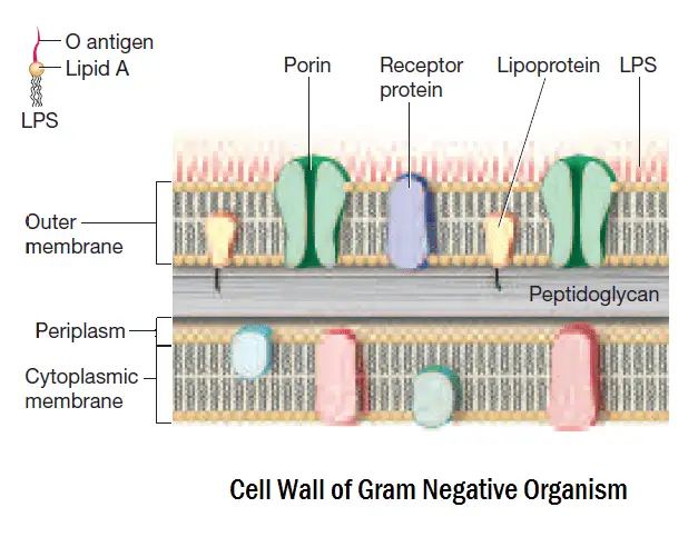
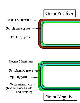
Gram Negative Cell Wall Diagram
Information from its description page there is shown below. It consists of a complex lipid with attached polysaccharide.
 A Diagram Of A Gram Negative Cell Wall B Electron Micrograph Of
320 160 pixels 640 320 pixels 1024 513 pixels 1280 641 pixels 1486 744 pixels.
Gram negative cell wall diagram. These bacteria have a wide variety of applications ranging from medical treatment to industrial use and swiss cheese production. E it is sensitive to penicillin. This is a file from the wikimedia commons.
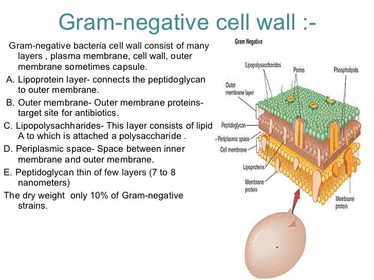
Filegram negative cell wallsvg. C it protects the cell in a hypertonic environment. 800 401 pixels.
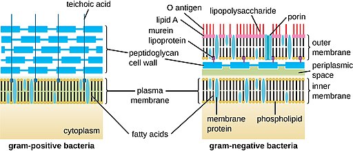
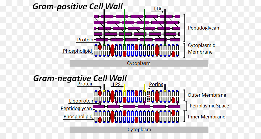
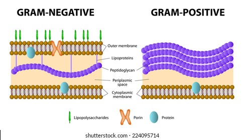
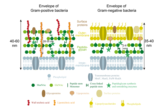
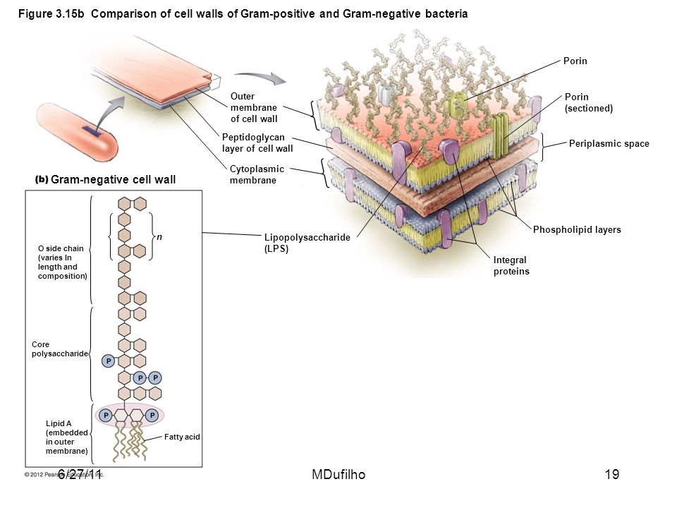
B it is sensitive to lysozyme. The cell wall of gram negative bacteria is thin approximately only 10 nanometers in thickness and is typically comprised of only two to five layers of peptidoglycan depending on the growth stage. In gram positive bacteria the cell wall is much thicker 20 to 40 nanometers thick.
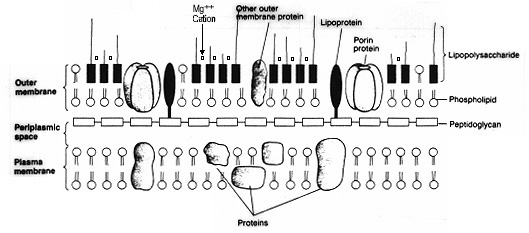
It is a complex structure with three components outside the peptidoglycan layer. Peptidoglycan makes up only 5 20 of the cell wall and is not the outermost layer. Peptidoglycan pep tid o gly can is a molecule found only in the cell walls of bacteria.
Size of this png preview of this svg file. Start studying gram negative cell wall. Amount and location of the peptidoglycan molecule in the prokaryotic cell wall determines whether a bacterium is gram positive or gram negative.
Gram negative cell walls are strong enough to withstand 3 atm of turgor pressure 40 tough enough to endure extreme temperatures and phs eg thiobacillus ferrooxidans grows at a ph of 15 and elastic enough to be capable of expanding several times their normal surface area 41. Prokaryotes with protection from the environment. The peptidoglycan of gram negative bacteria is located between the plasma membrane and an outer lps membrane.
Compared with gram positive bacteria gram negative bacteria are more resistant against antibodies because of their impenetrable cell wall. Learn vocabulary terms and more with flashcards games and other study tools. The cell walls of gram negative bacteria are more chemically complex thinner and less compact.
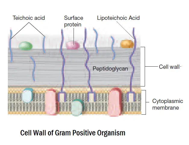
In figure 43 which diagram of a cell wall is a toxic cell wall smaller gram negative in figure 43 which diagram of a cell wall has a wall that protects against osmotic lysis. D it contains teichoic acids. 2 each of the following statements concerning the gram positive cell wall is true except a it maintains the shape of the cell.
A gram negative cell wall salmonella escherichia.
 Differences Between Gram Positive And Gram Negative Bacteria
 Cell Wall Bacterial Cell Structure Gram Negative Bacteria Gram


 Gram Positive Vs Gram Negative Diagram Lima Stanito Com
 Glycopedia
 Differences Between Gram Positive And Gram Negative Bacteria
 Gram Negative And Gram Positive Cell Walls Grampositive Bacteria
 Differences Between Gram Positive And Gram Negative Bacteria
 Differences Between Gram Positive And Gram Negative Bacteria
 Cell Wall Of Bacteria

 Schematic Structure Of Gram Positive And Gram Negative Cell Walls
 Cell Structure And Function Ppt Video Online Download

 My Scientific Blog Research And Articles The Bacterial Cell Wall
 Gram Positive Facts For Kids
Cell Wall Gram Positive Vs Negative Bacteria Easybiologyclass
0 Response to "Gram Negative Cell Wall Diagram"
Post a Comment